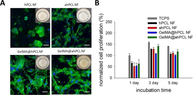
Figure 4.
In vitro cultivation of HDF with various NFs and spontaneous formation of HDF/NF cell sheets. (A) CLSM of HDF/NF cell sheets at day 3. Z-stacked images (10 slices per sample, slice thickness = 4.1–6.9 μm) were superimposed to show 3D-associated HDF/NF cell sheets stained for nucleus (blue) and F-actin (green). Insets are digital photos of HDF/NF cell sheets in cell culture plates. (B) Cell proliferation of HDF in NF for 5 days based on the WST-1-based cell viability assay. The absorbance at 450 nm of all samples was normalized with respect to that of cells in TCPS at day 1.

