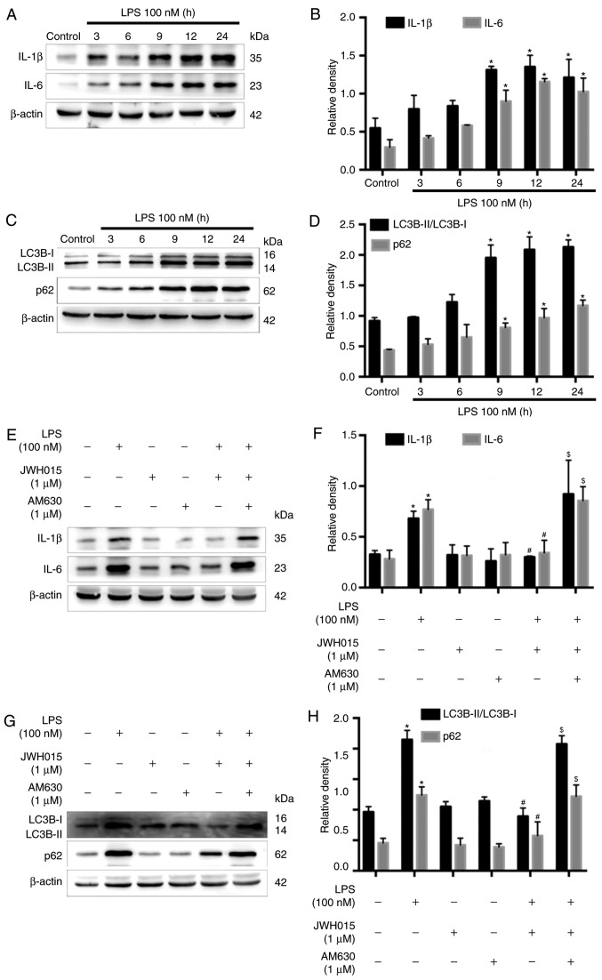
Figure 7.
Effects of JWH015 on inflammatory factors and autophagy in primary neurons. Primary neurons were stimulated with LPS (100 nM) for 0, 3, 6, 9, 12 and 24 h. (A) Western blotting demonstrated the upregulation of IL-1β and IL-6 after LPS-stimulation. (B) Quantification of IL-1β and IL-6 in the primary neurons after LPS-stimulation. *P<0.05 vs. Control (0 h). (C) Increased ratio of LC3B-II/LC3B-I and increased expression of p62 in LPS-stimulated primary neurons. (D) Quantification of LC3B-II/LC3B-I and p62. *P<0.05 vs. Control (0 h). (E-H) Primary neurons were treated with JWH015 (1 µM) and AM630 (1 µM) for 12 h. (E) Inhibition of IL-1β and IL-6 after treatment with JWH015 in LPS stimulated-primary neurons. (F) Quantification of IL-1β and IL-6 in different groups. *P<0.05 vs. control group; #P<0.05 vs. LPS group; $P<0.05 vs. LPS + JWH015 group. (G) Decreased ratio of LC3B-II/LC3B-I and decreased expression of p62 after treatment with JWH015 in LPS-stimulated primary neurons. (H) Quantification of LC3B-II/LC3B-I and p62 in different groups. *P<0.05 vs. control group; #P<0.05 vs. LPS group; $P<0.05 vs. LPS + JWH015 group. In all western blots, β-actin was used as a loading control; data are expressed as the mean ± SD; n=3. IL, interleukin; LC3B, microtubule-associated protein 1 light chain 3β; LPS, lipopolysaccharide.

