Fig. 3.
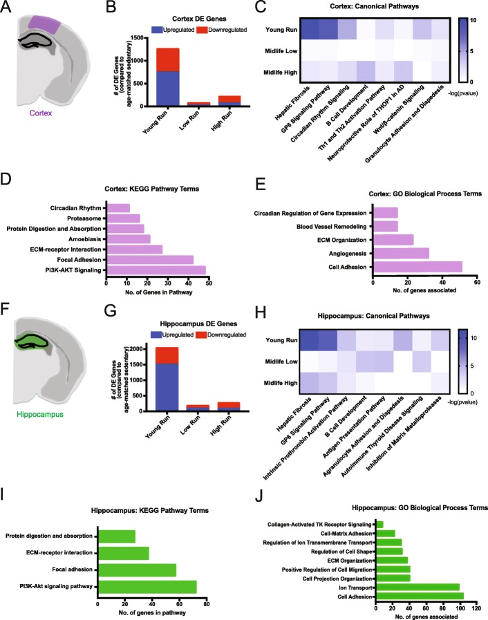
RNA-seq analysis identified ECM-related enrichment in both the cortex and hippocampus of young runners. a Cortical region (purple) used for RNA-seq. b Number of DE genes (FDR < 0.05) found in the cortex of the young runners compared to young sedentary (‘Young Run’), midlife low runners compared to midlife sedentary (‘Low Run’), and midlife high runners compared to midlife sedentary (‘High Run’) (FDR < 0.05). c IPA canonical pathway analysis showed enriched ‘Hepatic Fibrosis’ and ‘GP6 Signaling Pathway’ in the cortex of young run compared to young sedentary, however not significant in midlife comparisons. d KEGG pathway enrichment analysis in the cortex for young run compared to young sedentary (FDR < 0.05). e GO term enrichment analysis in the cortex for young run compared to young sedentary (FDR < 0.05). f Hippocampal region (green) used for RNA-seq tissue submission. g Number of DE genes (FDR < 0.05) found in the hippocampus of the young runners compared to young sedentary (‘Young Run’), midlife low runners compared to midlife sedentary (‘Low Run’), and midlife high runners compared to midlife sedentary (‘High Run’) (FDR < 0.05). h IPA canonical pathway analysis showed enriched ‘Hepatic Fibrosis’ and ‘GP6 Signaling Pathway’ in the hippocampus of young run compared to young sedentary, however not significant in midlife comparisons. i KEGG pathway enrichment analysis in the hippocampus for young run compared to young sedentary (FDR < 0.05). j GO term enrichment analysis in the hippocampus for young run compared to young sedentary (FDR < 0.05)

