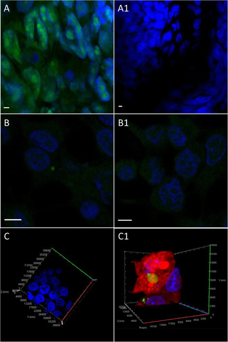
Fig. 2.

Visualization of 5-ethynyl uridine (EU)-labelled RNA in trophoblast spheroids and endometrial cells. a RNA in trophoblast spheroids were labelled with 5-ethynyl uridine (EU) and stained with Alexa azide. Green florescence is evidence of successful labelling. a1 Unlabelled spheroids (negative control) did not show fluorescent signal. b Endometrial cells were stained with Alexa azide after 24 h incubation with labelled spheroids to visualize the transferred transcripts. Green dots in endometrial cells indicate labelled RNA transfer. b1 Endometrial cells co-incubated with unlabelled spheroids were used as negative controls. Negative control did not exhibit any specific fluorescent signal. c, c1 3-dimentional confocal scanning of endometrial cells with cytoplasmic EU labelled RNA with and without cell tracker dye. Scale bar 4 μm
