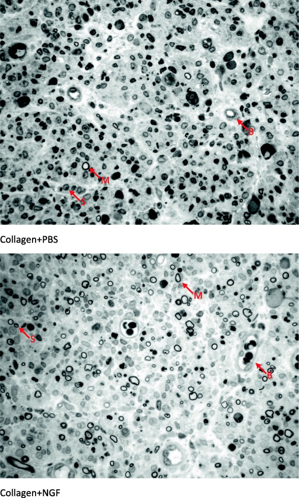
Fig. 7.

Light micrographs of representative cross sections in regenerated nerves treated with different conduit fillings. Myelin sheath (M) of the regenerated axon was stained dark blue with the toluidine blue. Blood vessels (B) and Schwann cells (S) are interspersed among these axons. Scale bar = 20 μm
