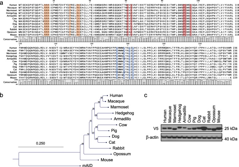
Fig. 1.
Comparison of APOBEC1 cytidine deaminases. a CLUSTALW alignment of A1 protein sequences. Residues involved in zinc coordination are depicted in red. Residues in orange are part of A1 bipartite nuclear localization signal while those involved in nuclear export of A1 are represented in blue. b Phylogenetic tree of A1 protein sequences constructed using the Neighbor-joining method with the CLC Main Workbench 7.0.2 software. Mouse AID was used to root the tree. Numbers correspond to bootstrap values inferred from 100,000 replicates. c Western blot analysis of V5-tagged A31 proteins in quail QT6 cells. β-actin probing was used as loading control

