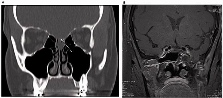
Figure 1.
A, Coronal CT sinus w/o contrast. Patient S. M., a 41-year-old woman with thinning of the cribriform plate and fovea ethmoidalis (arrow). Low-attenuation lesion representing the meningoencephalocele. B, Coronal MRI T1-weighted post contrast. Patient N. C., a 62-year-old woman with 8 mm defect in the floor of the left middle cranial fossa with herniating meningoencephalocele into the left sphenoid sinus.

