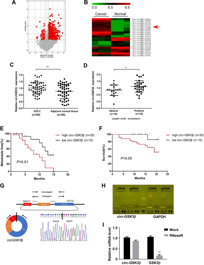
Fig. 1.
CircGSK3β was overexpressed in ESCC and correlates with poor patient prognosis. a Volcano plot compared the expression fold changes of circRNAs for ESCC tissues versus adjacent normal tissues. The red dots represented circRNAs with significantly changed expression level. b Clustered heatmap for top 20 upregulated and downregulated circRNAs, with rows representing circRNAs and columns representing tissues. The numerical data represented the serial number of circRNAs in circBase. c Scatter plots illustrating qRT-PCR analysis of expression fold change for circGSK3β in ESCC tissues compared with matched adjacent normal tissues. d Statistical analyses of circGSK3β expressions in different lymph node metastasis samples. e and f Statistical analyses of the association of circGSK3β expression with MFS (e) and OS (f) in ESCC patients. g Schematic illustration of circGSK3β locus with specific primers and Sanger sequencing result of circGSK3β. h RT-PCR products with divergent primers showing circularization of circGSK3β. i RT-qPCR analysis of circGSK3β and GSK3β transcripts in the presence or absence of RNase R treatment, respectively

