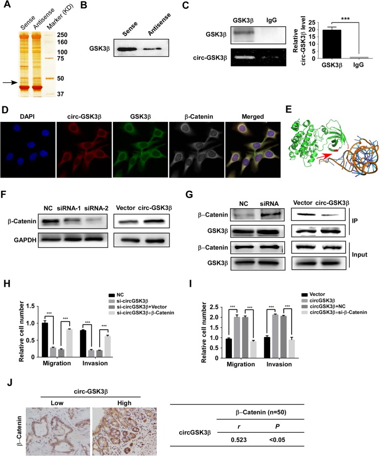
Fig. 3.
circGSK3β interacts with GSK3β and promotes metastasis by β-catenin. RNA pull-down experiment with ESCC cell lysate. The proteins were visualized by silver staining, and indicated spots were analyzed by mass spectrometry (a) or immunoblot of GSK3β (b). c QPCR detection of circGSK3β retrieved by GSK3β-specific antibody compared with immunoglobulin G (IgG) in the RIP assay. d RNA FISH assay of circGSK3β combined with immunofluorescence detection of GSK3β and β-Catenin in ESCC cells. e Graphical representation of three-dimensional structures of the interaction model of circGSK3β with GSK3β by SPOT-RNA. f β-catenin protein levels in ESCC cells with circGSK3β depletion or overexpression were detected by immunoblotting. g Immunoblot detection of indicated proteins in GSK3β-immunoprecipitated complex from lysates of ESCC cells with circGSK3β knockdown or overexpression. h and i ESCC cells with depletion of circGSK3β (Η) were transfected with β-catenin and circGSK3β-overexpressed cells (i) were transfected with control siRNA or siRNA against β-catenin. Migration and invasion abilities of ESCC cells were detected by Transwell and Matrigel invasion assay, respectively. j Statistical analyses of the association between circGSK3β and β-catenin expression in ESCC tissues. r, Pearson correlation coefficient; *P < 0.05, **P < 0.01, ***P < 0.001

