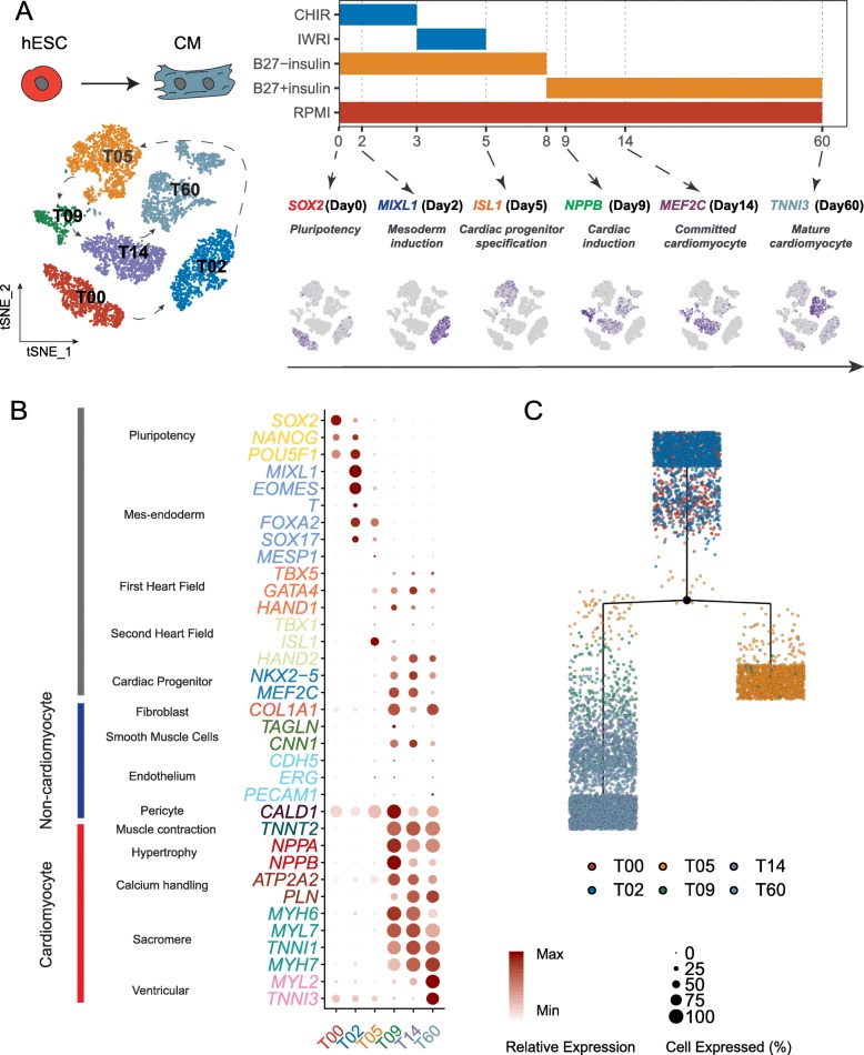
Fig. 1.
Comprehensive analysis of cardiac differentiation at single-cell resolution. a Schematic of the experimental design. Left: t-SNE plot of single-cell clustering from all six time points. Cells are colored by collected time points. Right: CM differentiation protocol (upper panel) and expression of selected markers at specific differentiation stages (lower panel). The shade of indigo color in the t-SNE plot reflects the relative expression level of corresponding genes. b The overall expression pattern of canonical markers representing the developmental stages/typical cell types at each time point. The size of the dots reflects the percentage of cells expressing specific marker, and the shade of the dots indicates the relative expression level. c Reconstruction of cell differentiation tree using top differentially expressed transcription factors of all time points. The black dot represents the bifurcation point observed on day 5. The end of the branch does not necessarily refer to a developmental end state

