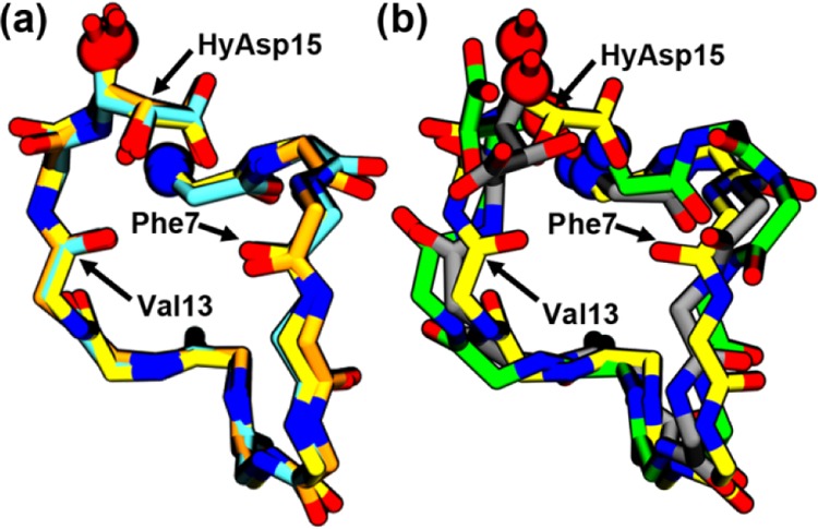
Figure 3.

Backbone alignment (aligned on Cα, N, C, and O atoms) of (a) the structures obtained after the equilibration of simulations Dflip (yellow), Dunflip (orange), and Eflip (cyan) is compared with (b) an alignment of the Dflip (yellow), Eunflip (green), and NMR (gray) structures. Only the nonhydrogen atoms of the backbone of residues 5–15 and the side chain of HyAsp15 are shown. The locations where the N- and the C-termini have been truncated from the images for clarity are shown as blue and red spheres, respectively.
