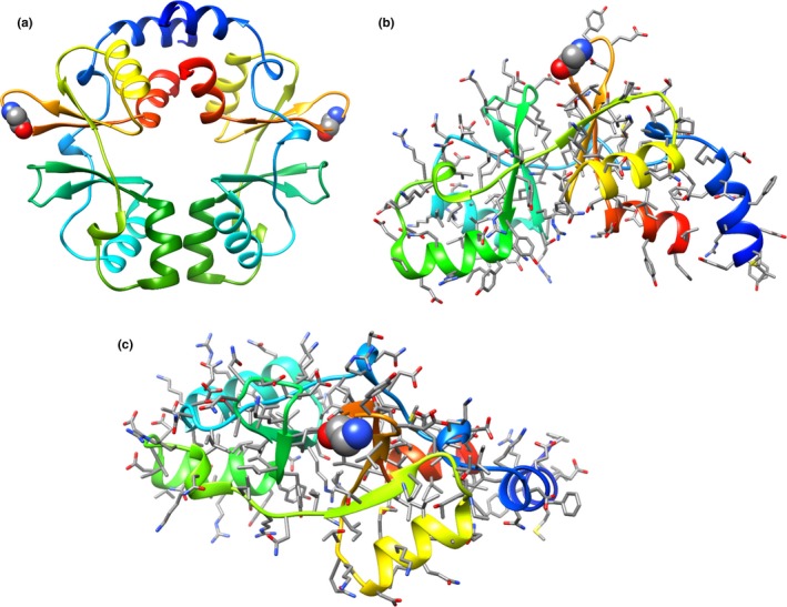
Figure 3.

Structural model of the LP965 CorC protein. (a) Top‐down view of proposed homodimer formed by tandem CBS domains. (b) Top‐down and (c) side‐views of (monomer) LP965 CorC protein region from Y216 to G351 shown in ribbon representation and colored by a rainbow scheme from N‐terminus region (blue) to C‐terminus region (red). All sidechains are shown in stick representation, except for G334, which is shown in space‐filling representation. Template‐based modeling was accomplished using PDB: 3OCO
