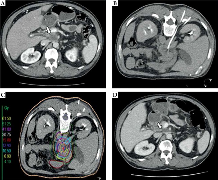
Fig. 1.
Interventional technique and local tumor control in a patient with a retroperitoneal lymph node metastasis (rLNM) from pancreatic ductal adenocarcinoma. A) Pre-interventional contrast-enhanced CT slice showing a rLNM (white arrow) located below the coeliac trunk; B) Peri-interventional CT slice with one percutaneously implanted brachytherapy catheter. The patient is placed in the prone position; C) Planning CT with indicated clinical target volume (CTV; blue line), isodose lines, and marked organs at risk (e.g. gastric and duodenal structures). The color-coded isodose levels are shown in Gy (scale on the left side of the image); D) Contrast-enhanced CT slice three months after high-dose-rate interstitial brachytherapy showing partial remission of the treated lesion

