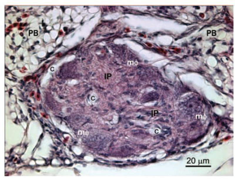
Figure 2.

Lung section from a 30-d-old broiler showing an arteriole with its lumen occluded by a plexiform lesion. The affected arteriole lies in the connective tissue septum separating 2 adjacent parabronchi (PB). The lesion consists of a matrix of proliferating intimal cells (IP) with foam-type macrophages (mΦ) arrayed around the remnants of the vascular wall. The perivascular connective tissue and gas exchange parenchyma contain nucleated avian erythrocytes and heterophils. The glomeruloid-like structure of this mature plexiform lesion is indicated by the multiple vascular channels (c) that have been cleared of erythrocytes by perfusion fixation. The 5-μm-thick section is stained with hematoxylin and eosin. Color version available in the online version of this paper.
