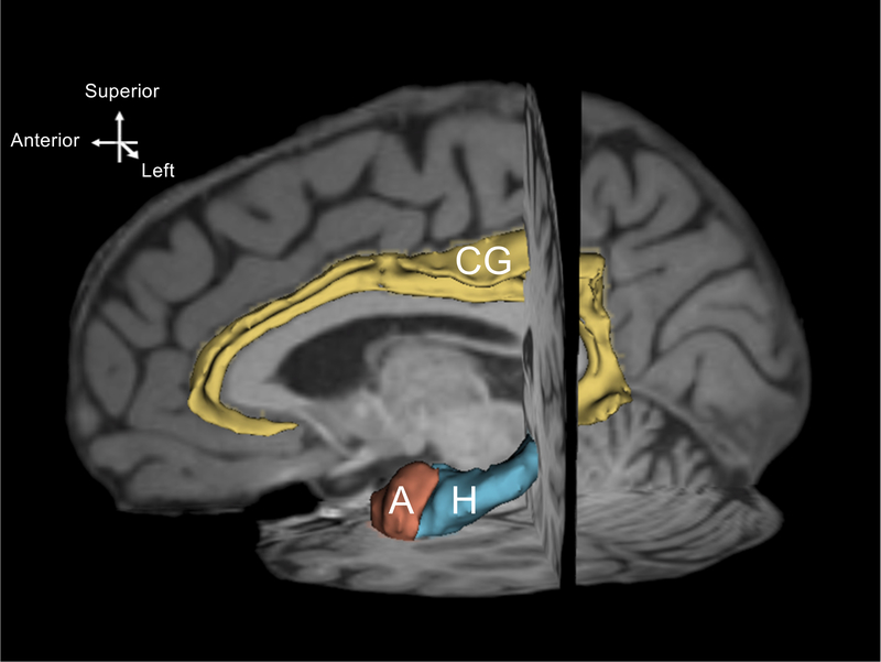
Fig. 1.
A three-dimensional reconstruction of left hemisphere regions of interest. The model was created from one randomly selected dataset using the model maker module of Slicer 4.1. Yellow = cingulate gyrus (CG), blue = hippocampus (H), terracotta = amygdala (A) The model is shown on a paramidsagittal slice and is superimposed on the individual T1-weighted images.

