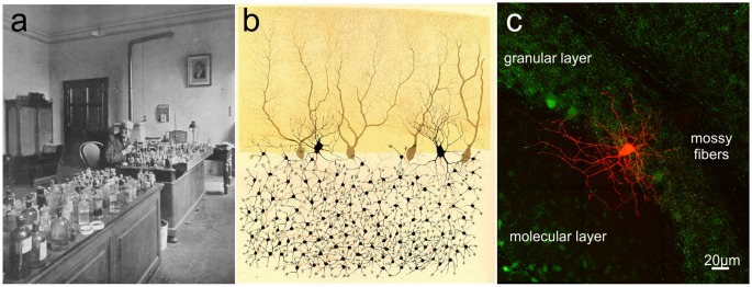
Figure 3.
Camillo Golgi and the cerebellar cortex. (A) Camillo Golgi in his laboratory at the University of Pavia. (B) Illustration by Camillo Golgi of a Golgi impregnated preparation of the cerebellum. Taken from Golgi (1883; available via license CC BY 4.0). (C) The current high-resolution rendering of a Golgi cell filled with a fluorescent dye and imaged with a two-photon microscope (courtesy of J. DeFelipe).

