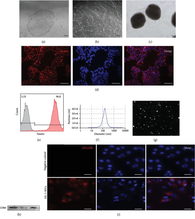
Figure 2.
Characterization of hESC-NSC and hESC-NSC-derived MVs. The phase morphology of hESCs and NSCs growing on Matrigel-coated dish (a, b). Typical morphological neurospheres of NSCs (c). hESC-NSC were immunofluorescence staining for nestin (d). Flow cytometry analyzed purity of nestin, gray line: isotype control; red line: hESC-NSCs (e). Nanoparticle trafficking analyzed the diameters of MVs. The particles were 1.17E + 10 particles/ml, and the diameters of the particles were within the range of 50–1000 nm, with the average of 152.5 nm (f). A representative screenshot of the NTA video, the bright white dot indicates one moving particle (g). Western blotting characterized microvesicle marker CD63, three replicate samples (h). Representative confocal microscopy of HL-1 cells that was exposed to Dil-labelled MVs (i). ((a–d): scale bars 100 μm; (h): scale bars 15 μm).

