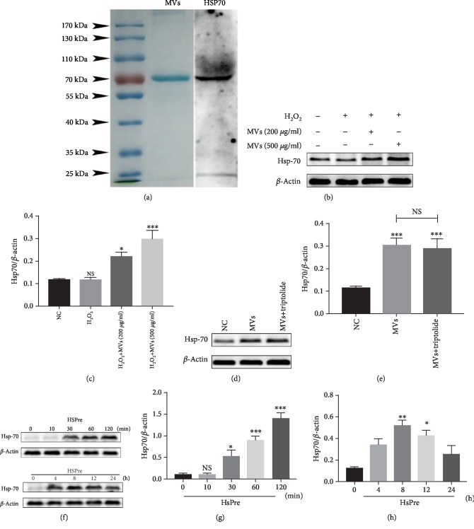
Figure 5.
Transfer of HSP-70 between cells via hESC-NSCs-derived MVs. Coomassie blue staining and western blot analysis of protein extracts from MVs (a). Western blot images showing the protein levels of the HSP-70 after HSPre with/without triptolide pretreatment (b–e). Western blot images showing the protein levels of the HSP-70 in HL-1 cells were exposed to 42°C for 10, 30, 60, and 120 min and then allowed to recover at 37°C for 0, 4, 8, 12, and 24 h. β-Actin was used as an internal control (f–h). Every experiment was repeated at least three times. Error bars indicate mean ± SD (∗P < 0.05; ∗∗P < 0.01; ∗∗∗P < 0.001).

