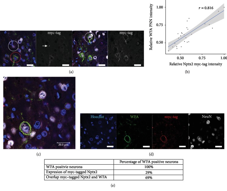
Figure 1.
Colocalization of myc-tagged Nptx2 and PNNs. 3-month-old rats were injected with lenti-cmv-Nptx2 into the somatosensory cortex to express myc-tagged Nptx2. Perfused tissue was cut in 20 μm sections and stained with 1 : 250 WFA to identify PNNs, 1 : 100 myc-tag antibody to identify the myc-tagged Nptx2 protein expressed by the lenti-cmv-Nptx2, and 1 : 200 NeuN antibody to identify neurons. (a) Representative images of the somatosensory cortex stained for WFA (green), myc-tag (red), and Hoechst (blue). In the left image, the white circle shows a neuron with intracellular myc-tagged Nptx2, but not on the cell membrane. The red circle shows a neuron with myc-tagged Nptx2 intracellularly and overlapping with the PNN. The white arrow points at the dotty myc-tag staining which is suggestive of vesicle staining. In the right image, the white circle shows a neuron which contains myc-tagged Nptx2 colocalizing with the PNN but shows no signs of intracellular myc-tagged Nptx2. (b) A correlation graph for the intensity of the myc-tag channel and the WFA channel is presented. A correlation analysis of WFA staining intensity and myc staining intensity revealed a correlation of 0.816 which indicates a high level of intensity correlation of the two channels in the PNN area. (c, d) The WFA staining for PNNs (green) is overlapping with the myc-tagged Nptx2 staining (red). (e) An overview of the number of neurons which are expressing the myc-tagged Nptx2 and which are positive for myc-tagged Nptx2 staining on their PNN. Intracellular myc-tag signal-positive neurons and PNN myc-tag signal-positive neurons were calculated. All images were taken in the somatosensory cortex (scale bar = 20 μm).

