Figure 1.
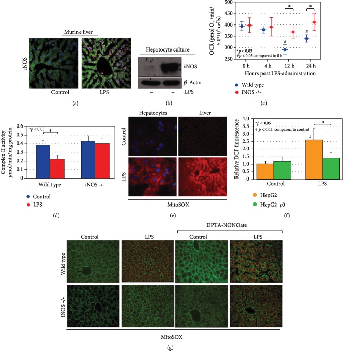
LPS induces mitochondrial dysfunction and mtROS production in an iNOS-dependent fashion. LPS induction leads to iNOS activation both in vivo (at 12 h exposure, (a)) and in cultured hepatocytes (following 6 h exposure, (b)). Cultured hepatocytes with prolonged exposure to LPS (times indicated) demonstrate a mitochondrial oxygen consumption defect over time that is dependent on iNOS expression (c). Evaluation of specific complex II activity in hepatocytes demonstrates a similar iNOS-dependent reduction of activity following LPS exposure (single time point, 12 h, (d)). In conjunction, LPS exposure is associated with increased mtROS production as measured by MitoSOX both in vivo and in vitro (12 h LPS exposure, 60 min preloading of MitoSOX, (e)). Global ROS production as measured by DCF fluorescence is predominantly mitochondrial in origin, as HepG2 ρ0 cells (which lack mitochondrial DNA) do not significantly generate ROS following LPS stimulation in comparison with HepG2 parent cells (12 h LPS exposure, (f)). MtROS generation following LPS administration as detected through in situ MitoSOX staining in liver sections is dependent on iNOS. MtROS production can be reconstituted in iNos−/− hepatocytes with the NO donor DPTA-NONOate, suggesting NO signaling downstream of iNOS is critical (12 h LPS and/or DPTA-NONOate, 60 min preloading of MitoSOX, (g)). In immunofluorescent imaging, green staining is from cytoskeletal actin as measured by 488-conjugated phalloidin antibody. Statistical significance is highlighted in individual panels as necessary.

