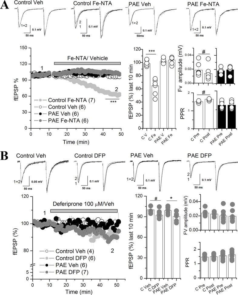
Figure 3.
Iron addition (Fe-NTA) and iron chelator (DFP) affects AMPAR synaptic transmission at the CA1 of the hippocampus of PAE rats compared to control rats. (A) Effect of Fe-NTA on basal transmission of PAE and control rats. Basal transmission (% fEPSP) was recorded on slices from control and PAE rats and 20 μM Fe-NTA or vehicle (Veh, V) was bath applied as is indicated in the graph. Bar graphs represent the average % fEPSP during the last 10 min, the fiber volley (Fv) amplitude and PPR pre HFS (Pre) and last 10 min of recording (Post) was represented in bar graphs. Note that FeNTA reduced basal transmission in control but not in PAE rats. Fv was decreased meanwhile PPR was larger (fEPSP2/fEPSP1) in control rats and not in PAE. (B) The effect of deferiprone (DFP) on basal transmission of PAE and control rats. After baseline, 100 μM DFP or vehicle (Veh, V) was bath applied as is indicated in the graph. Note that DFP incubation reduced basal transmission in PAE rats and controls. Fiber volley (Fv) amplitude and paired-pulse ratio (PPR) were not affected in PAE or control rats. In all panels, sample traces were taken at the times indicated by numbers on the summary plot; the number of litter is indicated in parentheses. ***p < value 0.0001, *p < value 0.05, repeated measured two way ANOVA followed Tukey’s post-hoc test. #p < 0.01 paired t test (A) and unpaired t test (B).

