Figure 4.

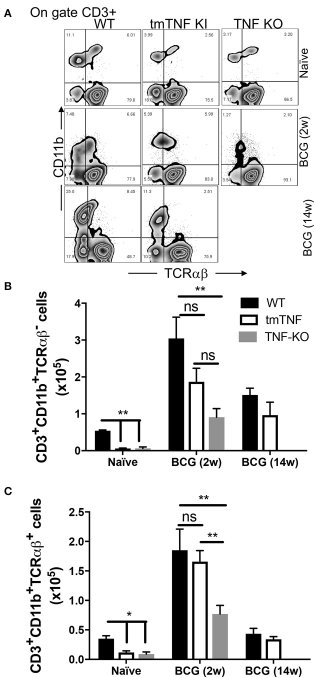
tmTNF regulates the presence of CD3+TCRαβ− and CD3+TCRαβ+ myeloid cells in the pleural cavity after a BCG infection. Wild type (WT), transmembrane TNF (tmTNF KI)-expressing and TNF knockout (TNF KO) mice were infected inside the pleural cavity with BCG. The animals were sacrificed at 2 weeks (BCG 2w) and 14 weeks (BCG 14w) post-infection, and a non-infected group (naïve) was used as the control. (A) Representative zebra plot: the CD3+ cells were delimited, and the co-expression of CD11b and TCRαβ were evaluated inside this gate in each group. The absolute numbers of CD3+CD11b+TCRαβ− (B) and CD3+CD11b+TCRαβ+ (C) subpopulations are shown; these numbers are obtained by measuring the relation between the percentage of cells (obtained by flow cytometry) with the total number of cells recovered from the pleural cavity. The data are expressed as the mean ± SD of n = 4–8 mice, three independent experiments. A statistical analysis was performed by two-way ANOVA with multiple comparisons, following by a Tukey test. **p < 0.01; ns = not statistic difference.
