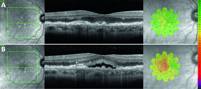
Figure 5.
Representative finding from a 64-year-old female study participant who developed SRF centrally in their right eye. (A) NIR scout image, SD-OCT B-scan, and MAIA findings 6 months prior to development of SRF. (B) NIR, SD-OCT, and MAIA at the time of SRF detection. Microperimetry scale of decibel values and corresponding colors is shown on the right.

