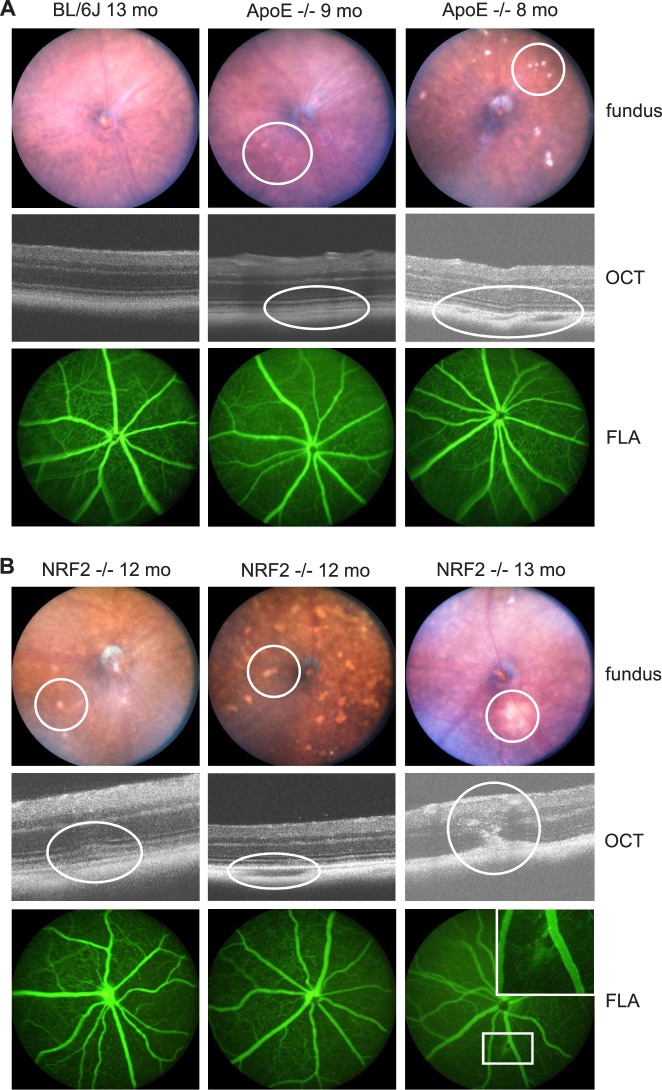
Figure 1.
Phenotype. (A) depicts a wild-type 13-month-old BL/6J mouse (left column) showing no AMD-like fundus alterations neither funduscopically (above), nor by OCT (middle), nor by FLA (below). Eight months (pre-SRT), as well as 9-month-old (1 month post SRT) ApoE−/− mainly showed mild AMD-like alterations (middle column) and rarely a phenotype of increased AMD-like alterations (right column). At the day of enucleation, 1 month after SRT, hence 9-month-old animals, there were no visible laser-induced alterations. DRS, as shown in the right column, funduscopically looked like drusen. OCT revealed, though, that they were mostly an area of hypopigmented, gap-like choroid adjacent to BrM. (B) Twelve-month-old (pre SRT), as well as 13-month-old (1 month post SRT) NRF2−/− mice showed a more severe AMD-like phenotype than ApoE−/−. Prominent DRS (left column), hypopigmented DRS-/atrophy-like areas (middle column) and in three eyes CNV formation connected to multiple DRS (right column) were typical for the NRF2−/− phenotype. In FLA images, CNV could be confirmed, if present. However, neither DRS nor possibly laser-induced hyperfluorescent spots were visible in FLA images.

