Figure 4.
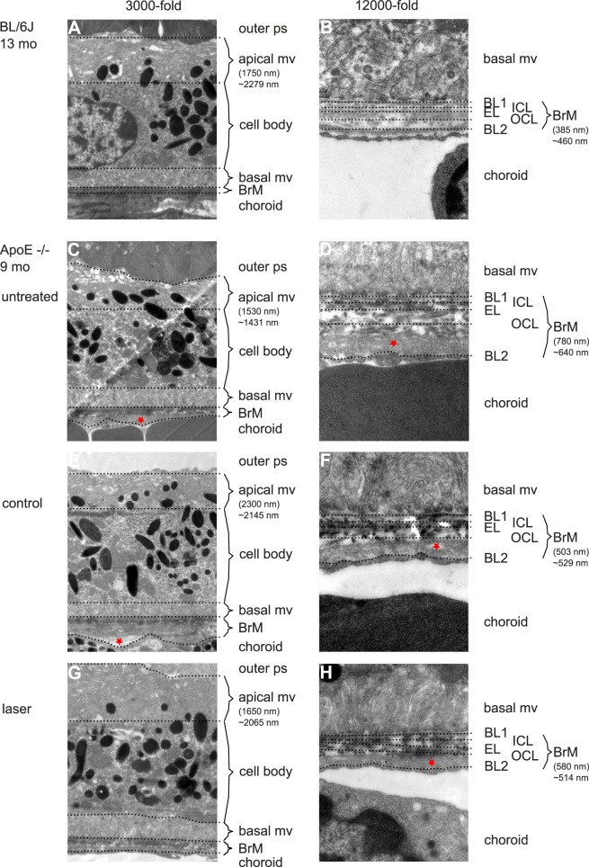
Secondary endpoint RPE morphology. (A) Three thousand-fold magnification of BL/6J RPE at 13 months of age. Outer photoreceptor segments (ps) are adjacent to RPE's apical mv. Length of bracket indicates the measured length at the given point. ∼ represents the mean value of the given eye. Note how variable thickness determination may be depending on the point of measurement. The cell body is characterized by a homogenous cytoplasm, the centered nucleus, mitochondria, inclusion bodies, and numerous melanosomes. Basal mv connect to BrM, which is a thin, multilayered mesh between RPE and choriocapillaris (choroid). (B) Twelve thousand-fold magnification of BrM of the same animal (B). Note the five layers of BrM. The basal mv of RPE connect to the first layer, RPE's basal lamina (BL1). Then three layers of collagen fibers form the mesh-like character of BrM, inner collagenous layer (ICL), elastic layer (EL), and outer collagenous layer (OCL). The basal lamina of choriocapillaris (BL2) as the fifth layer connects to the fenestrated choroidal endothelium. Bracket indicates the thickness measured at the given point. ∼ represents the mean thickness of the given eye. (C) Untreated 9-month-old ApoE−/− RPE. Note the reduced and morphologically altered apical mv and outer collagenous layer deposits (asterisk in [D]). (E and F) Display the control eye of a lasered mouse (asterisk indicates lipid-like deposits, typical for ApoE−/− mice). (G and H) Display the SRT-treated eye of the same mouse. Note the difference in BrM thickness between the lasered (H) and control eye (F) as well as to the untreated eye (D).

