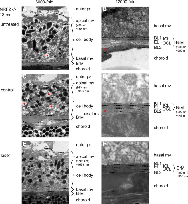
Figure 5.
Secondary endpoint RPE morphology. NRF2−/− mice showed a much more altered ultrastructural phenotype. Figure's presentation mode is analogous to Figure 4. Thickness measurements: brackets indicate the thickness measured at the given point, ∼ indicates mean of the given eye. (A) untreated NRF2−/− 13 months of age. Apical mv are shortened and morphologically altered/unorganized. Cytoplasm looks inhomogeneous, asterisk indicates a vacuole-like alteration of cytoplasm. BrM is variable in diameter and shows large outer collagenous layer deposits (B). (C) Control eyes show a similar phenotype; however, with reduced BrM thickness (D) compared with treatment naïve mice. (E) Morphologically SRT-treated RPE looks more organized and becomes similar to wild-type BL/6J RPE 1 month after treatment. (F) BrM is thinner compared with untreated mice. Thickness given in brackets only shows the size of the bracket. Measurements of BrM thickness need a standardized multimeasurements blinded system, because of variation in thickness even within one picture.

