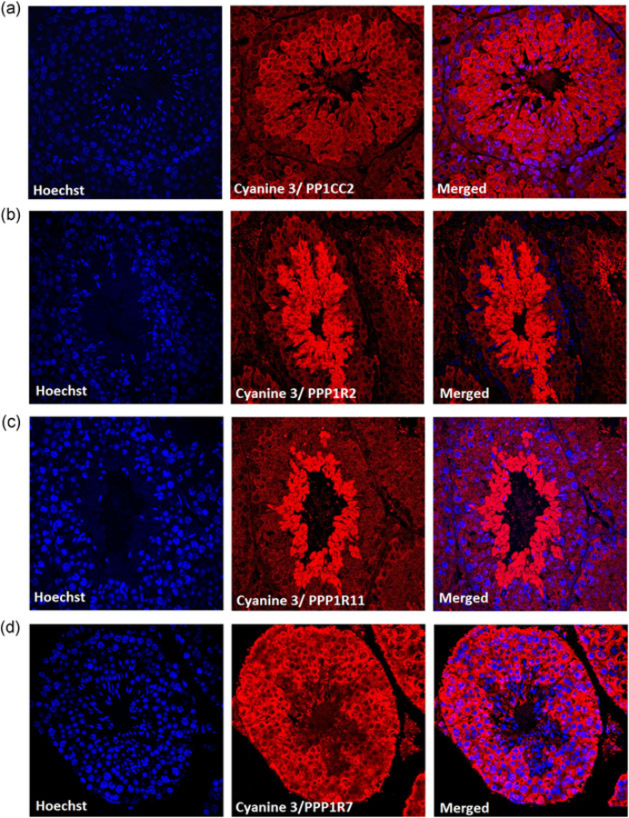
FIGURE 4.
Distribution of PP1γ2 and its binding partners in testis section. Immunohistochemistry of wild-type testis section show (a) PP1γ2 expression from secondary spermatocytes cytoplasm, spermatids, and mature spermatozoa. (b) PPP1R2 and (c) PPP1R11 is expressed in round spermatids and mature spermatozoa whereas (d) PPP1R7 is expressed throughout the cytoplasm of primary spermatocyte secondary spermatocyte round spermatid and mature spermatozoa. Cyanine 3 was used as a secondary antibody to visualize the proteins. Hoechst dye was used to stain the nucleus

