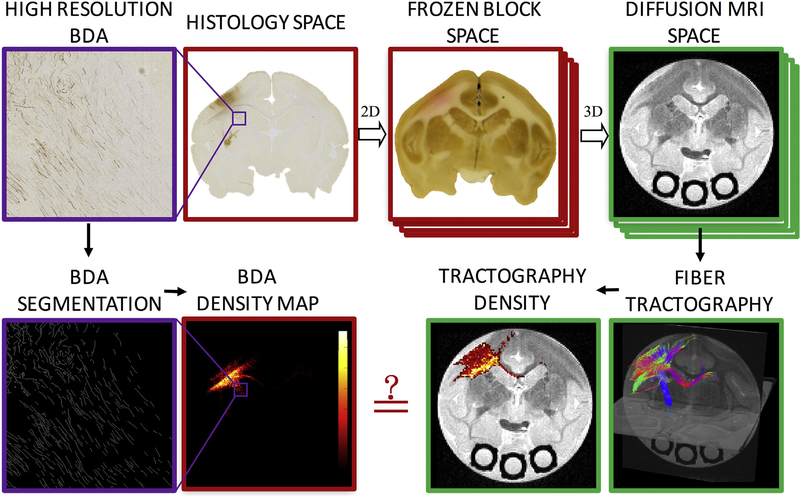
Fig. 1.
Methodology pipeline. High resolution BDA micrographs are registered to the corresponding digital photograph of the frozen tissue block, which is registered to the 3D diffusion MRI volume. From the micrograph, BDA is automatically segmented, resulting in a BDA density map. From diffusion MRI, tractography is performed, resulting in tract density maps. Direct, voxel-by-voxel comparisons can now be made between histology and diffusion tractography.

