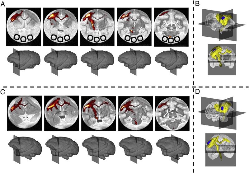
Fig. 2.
Histological Results. BDA density is shown overlaid on the non-diffusion weighted volume for five coronal slices for monkey #1 (A) and monkey #2 (C). A BDA mask is shown as a volume rendering indicating the presence of BDA in a given voxel for monkeys #1 (B) and #2 (D). Injection region is shown in blue, BDA mask is shown in yellow.

