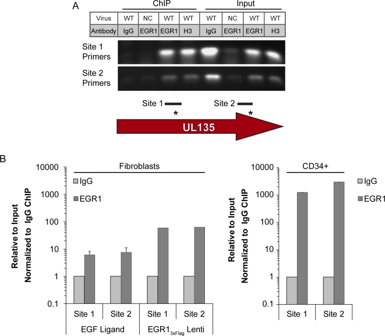
Fig 5. EGR1 transcription factor binds within the UL135 gene region.
(A) Fibroblasts were transduced with EGR13xFlag lentivirus and then infected with WT or UL133/8null mutant (negative control; NC) TB40/E virus (MOI = 1). Chromatin was immunoprecipitated (ChIP) with IgG control or antibodies specific to EGR1 or histone 3 (H3) and the presence of Site 1 or Site 2 was detected in the precipitates by PCR. As a positive control, PCR was also performed on 2% of the ChIP input. Gel is a representative experiment from 3 replicates. Diagram represents the amplicon region used for Site 1 and Site 2 detection. (B) ChIP-qPCR using SimpleChIP Enzymatic Chromatin IP Kit (Cell Signaling) was performed on fibroblasts infected for 48 h and pulsed with EGF for 1h, fibroblasts expressing EGR13xFlag infected for 48 h, or pure population of infect CD34+ HPCs in long-term culture for 5 days (6 dpi total). Fibroblasts were infected at an MOI of 1 and the CD34+ HPCs were infected at an MOI of 2. The presence of EGR1 Site 1 or Site 2 sequence was quantified by qPCR relative to a 2% input control and normalized to WT levels.

