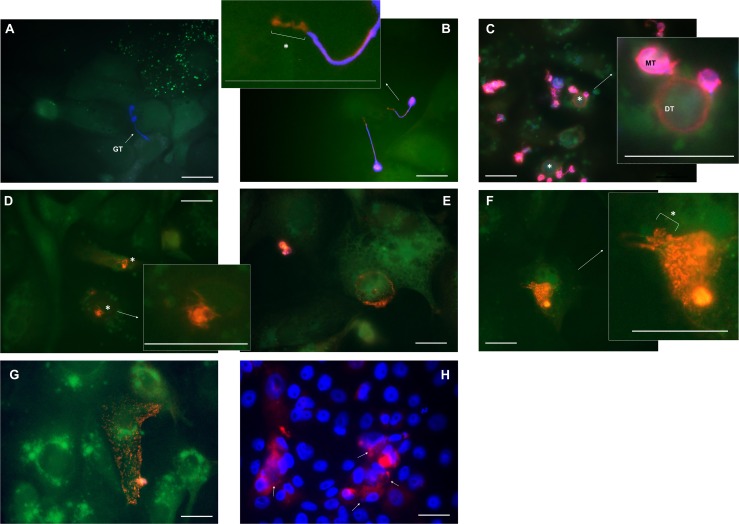
Fig 3. Bd development in A6 cells.
Shown is an overlay of the fluorescent signals of (A-G) Bd-infected A6 cells (green cell tracker), extracellular Bd (Calcofluor White (blue)) and extra-and intracellular Bd (Alexa Fluor 568 (red)) or (H) caspase-3 activation (red) and nuclear content (Hoechst (blue)). (A) Four hours after inoculation, formation of germ tubes (GT) was observed and (B) within 24 hours, these tubular structures penetrated the A6 cells (*). (C) At day 1–2 p.i., new intracellular chytrid thalli (*) are formed and the cell content of the mother thallus (MT) is transferred into the new daughter thallus (DT). (D) At day 2–3 p.i., the emptied mother thallus evanesces, resulting in intracellular Bd bodies (*) that (E) develop intracellularly into sporangia at day 3–4 p.i. (F) Once the sporangia reach the stage of a mature zoosporangium (day 4–5 p.i.), they use a discharge tube (*) to release their contents into the A6 cells (G). (H) At day 5–6 p.i., caspase-3 activation was observed in A6 cells associated with Bd (white arrow). Scale bar = 20 μm. Individual pictures of the different fluorescent channels can be found in S2 and S3 File.

