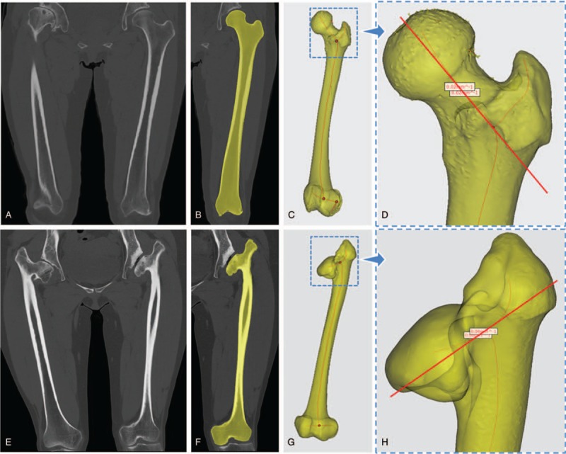
Figure 2.

Femoral nack axis determined by centerline and curvature. A, E: CT images of normal volunteer and DDH patient. B, F: mask of the femur separated from muscles, liagments and soft tissues. C, G: femoral centerline in the mode of transparency. D, H: points with minimal curvature to determin femoral neck axis.
