
Figure 2.
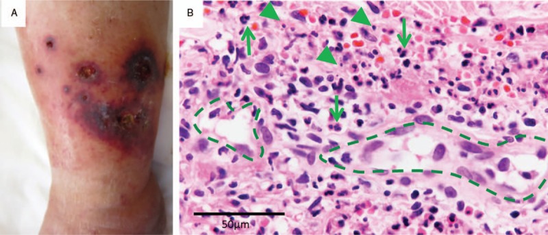
Skin findings. (A) Left ankle with purpuric lesions. (B) Light microscopy of the skin biopsy. Many neutrophils (arrow) infiltrated around vessels (dashed line) with nuclear fragments (arrowhead) (600×).
Official websites use .gov
A
.gov website belongs to an official
government organization in the United States.
Secure .gov websites use HTTPS
A lock (
) or https:// means you've safely
connected to the .gov website. Share sensitive
information only on official, secure websites.
Skin findings. (A) Left ankle with purpuric lesions. (B) Light microscopy of the skin biopsy. Many neutrophils (arrow) infiltrated around vessels (dashed line) with nuclear fragments (arrowhead) (600×).