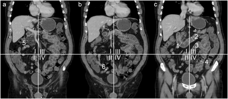
Fig 3. Coronal reformatted CT (three consecutive slices (a-c)) for demonstration of small bowel segment definition.
The abdomen is divided into 4 quadrants with the umbilicus as the center point. Representative measurements of the descending (1) and horizontal duodenum (2). Jejunal and ileal segments were defined using the 4-quadrant model: I = upper right quadrant, including the proximal ileum (5); II = lower right quadrant, including the distal ileum (6); III = upper left quadrant, including the proximal jejunum (3); IV = lower left quadrant, including the distal jejunum (4).

