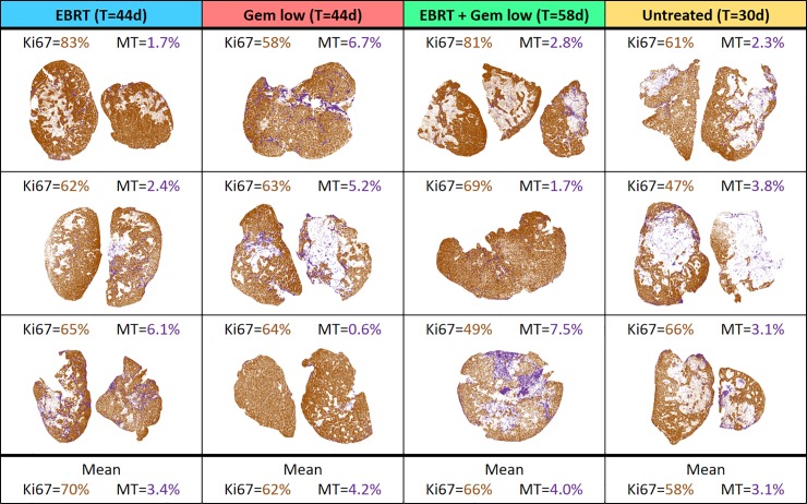
Fig 4. Overview of the results of the quantitative IHC analyses.
Analyses were performed for the animals treated with 5 Gy external beam radiotherapy (EBRT) and/or 60 mg/kg gemcitabine (Gem low) as well as for the untreated control animals. Quantification was made for digitalised histological sections stained with Ki67 (shown in brown) and Masson’s trichrome (MT, shown in purple). For each tumour, the percentage of the area positively stained for Ki67 (in viable tumour only) and MT (in the whole tumour area) was determined. Included is also the follow-up time, T, for each group.

