Figure 12.
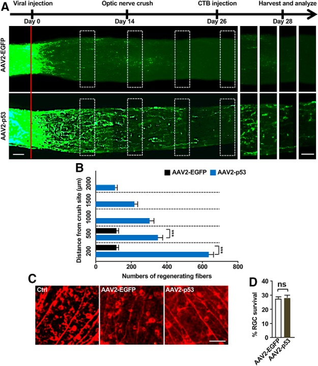
Overexpression of p53 enhances optic nerve regeneration. A, Top, Time line of the experiment. Bottom, Representative images showing that p53 overexpression in RGCs induced drastic optic nerve regeneration 2 weeks after the optic nerve crush. The right eight columns show enlarged images of nerves at places marked by white boxes on the left. The red line indicates the crush sites. Scale bars, 50 μm. B, Quantification of regenerating optic nerve axons at different distances from the nerve crush site (n = 6 nerves from 6 mice for each condition; p = 0.0001 at 200 μm, p = 0.0001 at 500 μm). C, Representative images of whole-mount retina stained with the neuron-specific βIII tubulin antibody Tuj-1. Scale bar, 100 μm. D, Quantification of Tuj-1-positive cells in C showing that overexpression of p53 did not affect RGC survival 2 weeks after optic nerve crush. ns, No significant difference. Data are represented as mean ± SEM. ***p < 0.001.

