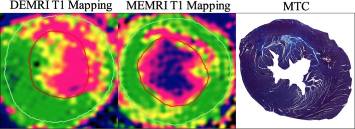
Figure 1.
Preclinical manganese-enhanced MRI (MEMRI). MEMRI T1 mapping of acute myocardial infarction in a preclinical model of myocardial infarction in rat, with MnDPDP. Colour maps are configured to show infarct (pink) and remote myocardium (green) with an intermediate peri-infarct zone (yellow). Note the visibly smaller infarct size with MEMRI compared with DEMRI, more in line with histological description of infarct with Masson’s trichrome (MTC). DEMRI, delayed-enhancement MRI; MnDPDP, manganese dipyridoxyl diphosphate.

