. 2019 Nov 13;8:e46302. doi: 10.7554/eLife.46302

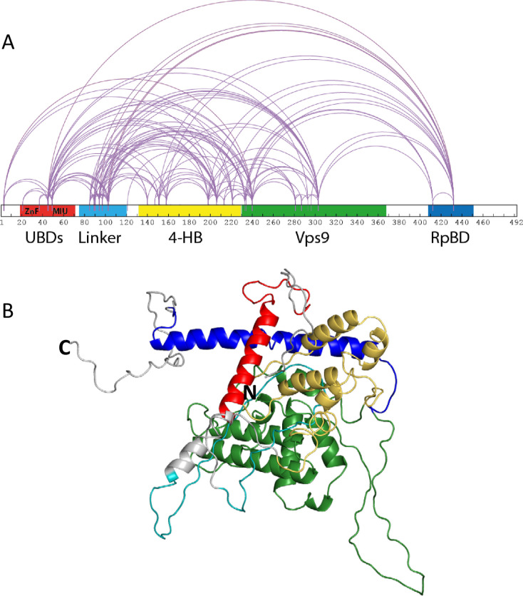
Figure 1. Rabex5 cross-linking-MS data and structural model.

(A) The cross-linking-MS data are shown for apo Rabex5 and illustrated in Xwalk (Kahraman et al., 2011). Ubiquitin binding domains (both ZnF and MIU together in red), Linker (teal), 4-helical bundle (gold), Vps9 domain (green), and rabpatin5 binding domain (blue). (B) A structural model of apo Rabex5 is pseudo-colored as illustrated above. This model is one of the possible arrangements and was chosen because it was in best agreement with the cross-linking-MS and HDX-MS data.

Figure 1.

Figure 1—figure supplement 1. Alignment and superimposition of our model with 1TXU.
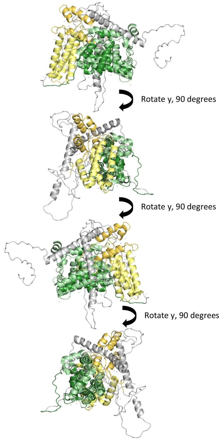
Figure 1—figure supplement 1.

Our best model was aligned and superimposed with the coordinates from 1TXU to illustrate relative positions between the 4-HB and Vps9 domain. Please keep in mind that the model we use for illustration is one of many possible models created herein. As with all static pictures, it does not capture the true ensemble of conformations.

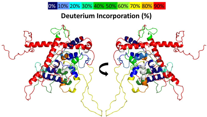
Figure 1—figure supplement 2. Deuterium uptake in Rabex5.

Figure 1—figure supplement 2.

The results of 10 s deuterium uptake are shown for WT apo Rabex5, using the indicated color scheme. The region in white has no peptide coverage.