Figure 3.

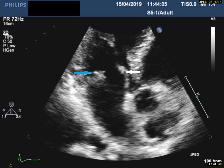
Focused view of the mitral valve in the apical three-chamber view demonstrating echogenic mass on the posterior leaflet (indicated by the blue arrow) and thickened anterior leaflet (indicated by the white arrow).

Focused view of the mitral valve in the apical three-chamber view demonstrating echogenic mass on the posterior leaflet (indicated by the blue arrow) and thickened anterior leaflet (indicated by the white arrow).