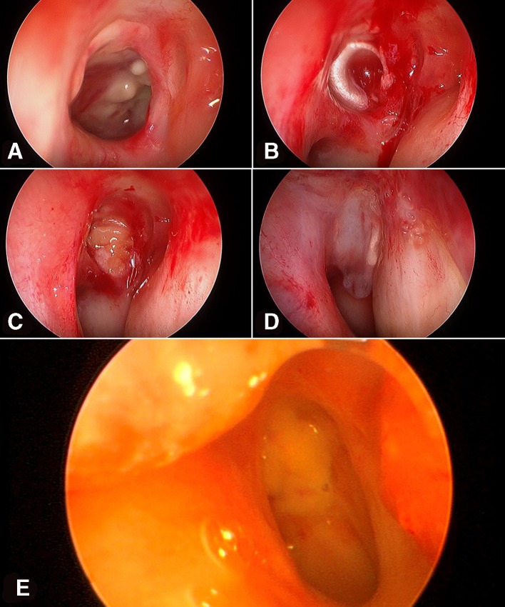
Figure 2.
The second surgical procedure. (A) Intraoperative endoscopic view shows the regression of the inflammatory signs with the persistence of a deep ulcer of the nasopharynx on the left side. (B) Endoscopic view shows the application of the first layer of fascia lata in the deep part of the cavity of the ulcer. (C) Endoscopic view shows the application of the fat grafts to filled the cavity of the ulcer. (D) Endoscopic view shows the application of the second layer of fascia lata on the surface of the mucosa of the posterior wall of the nasopharynx. (E) Endoscopic view 4 months after the surgery shows a good healing and integration of the grafts with a compete closure of the defect of the nasopharynx on the left side.

