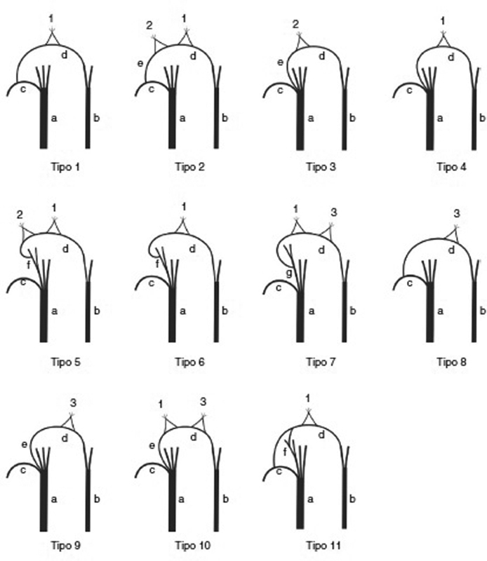
Fig. 9.

Schematic drawing of the different patterns of the Riché-Cannieu anastomosis. ( 1 ) Branches to the deep head of the FPB muscle; ( 2 ) branches to the superficial head of the FPB; ( 3 ) branches to the adductor muscle of the thumb. ( a ) Median nerve; ( b ) ulnar nerve; ( c ) recurrent branch of the median nerve; ( d ) deep branch of the ulnar nerve; ( e ) branch isolated from the main trunk of the median nerve; ( f ) digital radial collateral branch of the thumb; ( g ) first common digital branch of the median nerve.
