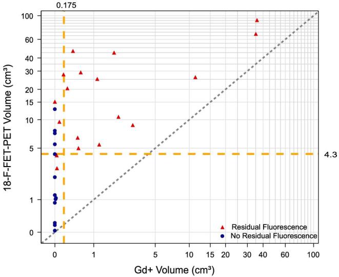
FIGURE 3.

Scatter plot of Gd+ and 18F-FET-PET volumes of each patient. Dotted bisectrix refers to perfect matching absolute values of Gd+ and 18F-FET-PET volumes. Intersected lines at x and y axes refer to cut-off values of 0.175 cm3 for Gd+ (indicating a “complete” resection by MRI standards) and 4.3 cm3 for 18F-FET respectively, the cut-off found for survival with this modality. For the group with no residual fluorescence median Gd+ volume was 0 cm3 (IQR 0-0) and median 18F-FET-PET volume was 1.2 cm3 (IQR 0.05-6.08). In the group with residual fluorescence Gd+ volume was 0.55 cm3 (IQR 0.15-2.34) and 18F-FET-PET volume was 17.82 cm3 (IQR 6.25-33.14). The graph indicates “complete” resections based on visible fluorescence to extend well into the residual 18F-FET-PET volume.
