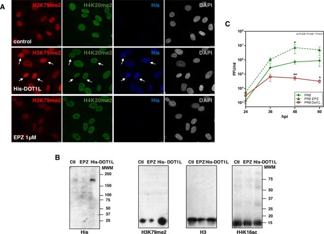
Figure 2.
A549 untransfected cells (control), transfected with plasmid His-Dot1L 48 h (His-DOT1L) or treated with pinometostat 48 h (EPZ) were used (A) for immunofluorescence analysis against (His), H3K79me2 and H4K20me2. White arrows denote cells with high expression of His-DOT1L. (B) Western blots against His-DOT1L, H3K79me2, H3 and H4K16ac. (C) Untransfected cells (PR8), transfected with His-Dot1L 48 h (PR8 Dot1L) or treated with pinometostat 48 h (PR8 EPZ), were infected with 10−3 pfu/cell with PR8 influenza virus and infective particles analyzed by plaque assay. Three independent experiments were performed. ns P > 0.05; *P < 0.05; **P < 0.01; ***P < 0.001.

