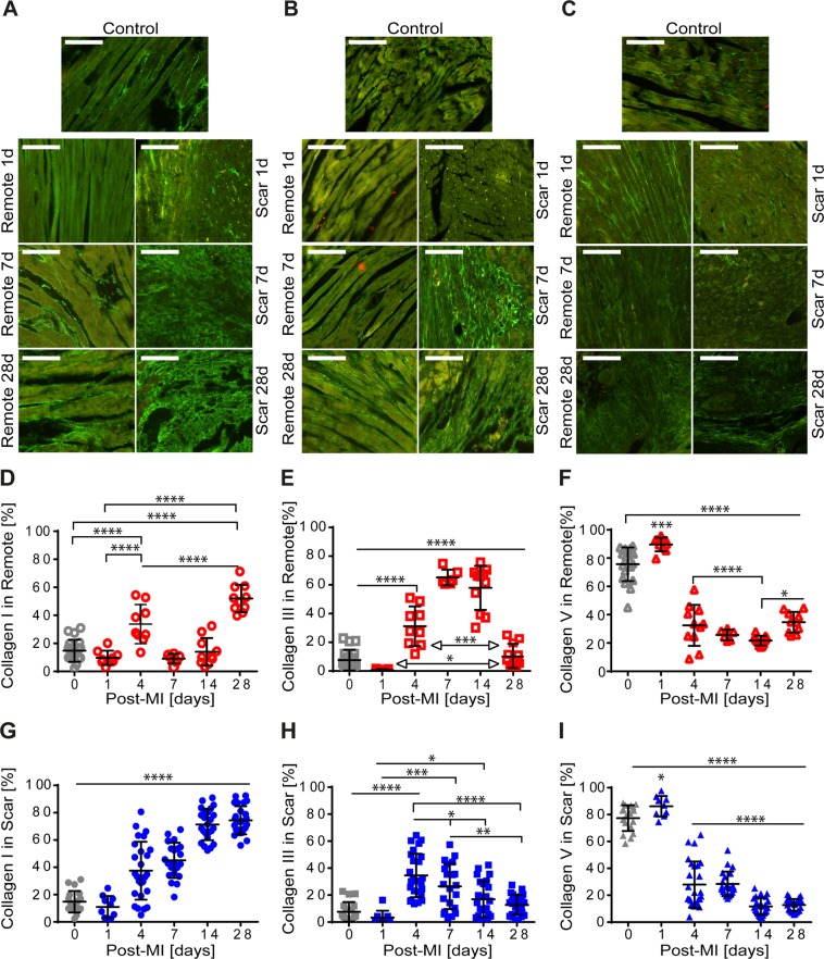
Figure 3.
Immunohistological characteristics of types I, III, and V collagen in control and remote vs. scar within the healing period of 28 days post-MI. Spatial distribution of type I collagen in remote (A-left) and scar (A-right), type III collagen in remote (B-left) and scar (B-right), and type V collagen in remote (C-left) and scar (C-right) control (upper panels). Scale bar: 50 μm. Time variation of the amount of types I, III, and V collagen in remote (D–F) and in scar (G–I). The collagen amount is normalized by the total amount of type I, III, and V collagen in remote and scar. (*p < 0.5, ***p < 0.001, ****p < 0.0001).

