Figure 1.

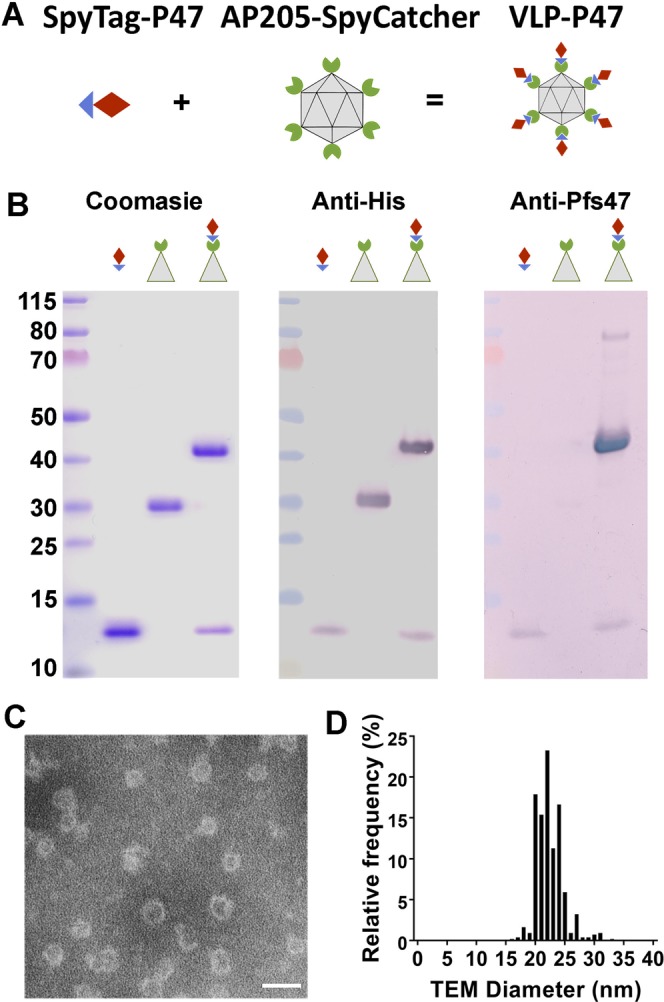
AP205-SpyCatcher and SpyTag-P47 isopeptide bond formation. (A) Schematic representation of the AP205-SpyCatcher and SpyTag-P47 isopeptide bond formation. Diagrams show SpyCatcher in green, Spytag in blue, and P47 in red. (B) Coomassie blue staining of SpyTag-P47, AP205-SpyCatcher, and conjugated VLP-P47 in SDS-PAGE after boiling and reducing in SDS-loading buffer (left). Anti-his western blot of SpyTag-P47, AP205-SpyCatcher, and conjugated P47-VLP (center). Anti-Pfs47 western blot of SpyTag-P47, AP205-SpyCatcher, and conjugated VLP-P47 (right). (C) TEM of VLP-P47 after negative staining with 2% uranyl acetate. (D) Size distribution of VLP-P47 from TEM image (n = 559). The average hydrodynamic diameter is 22.48 +/− 2.26 nm. Scale bar: 50 nm.
