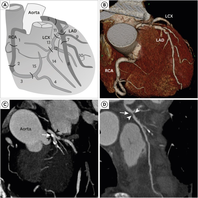
Fig. 1. The anatomy of the coronary arteries and the representative CCTA images of coronary artery disease. (A) A schematic drawing of the anatomy and segmentation of the coronary arteries. Fifteen segments are as follows: 1 - proximal RCA; 2 - middle RCA; 3 - distal RCA; 4 - posterior descending; 5 - left main; 6 - proximal LAD; 7 - middle LAD; 8 - distal LAD; 9 - diagonal 1; 10 - diagonal 2; 11 - proximal LCX; 12 - ob marginal; 13 - distal LCX; 14 - ob marginal 2; 15 - posterolateral branch. (B) Three-dimensional reconstruction of CCTA image in a RAO patient. (C) CCTA showed 20%–30% of stenosis (arrow heads) at left main coronary artery due to calcified plaque (arrow) in a 68-year-old man with central RAO. (D) CCTA showed severe stenosis (arrow heads) at proximal LAD due to non-calcified plaque (arrow) in a 72-year-old man with central RAO.
RAO = retinal artery occlusion, CCTA = coronary computed tomographic angiography, RCA = right coronary artery, LCX = left circumflex artery, LAD = left anterior descending.

