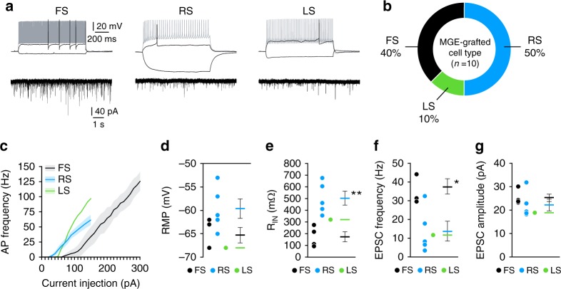
Fig. 2.
Transplanted MGE cells integrate into brain injured hippocampus as mature interneurons. a Voltage responses of three transplanted MGE cells to a hyperpolarizing current pulse (−50 pA) and depolarizing current pulses near threshold (black) and maximal firing (gray). Holding potential was near −70 mV. FS fast spiking, RS regular spiking, LS late spiking. Recordings were obtained from slices 45–60 DAT. Voltage-clamp recordings of EPSCs in each cell are shown below each current-clamp recording. b Occurrence of each interneuron subtype recorded based on firing properties (n = 10 cells from 3 TBI-MGE mice). c Plot of action potential firing frequency (Hz) as a function of current intensity. d Quantification of resting membrane potential (RMP) for each cell type. e Quantification of Rinput for each cell type. **P = 0.004, FS vs RS, two-tailed t-test. f, g Mean EPSC frequency (f) and amplitude (g) according to interneuron subtype. *P = 0.01, FS vs RS, two-tailed t-test. Error bars, s.e.m. Source data are provided as a Source Data file

