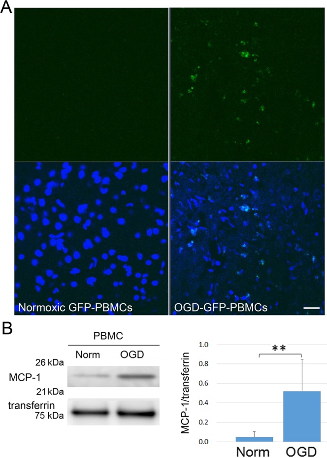
Figure 2.

The migration of administrated OGD-PBMCs. (A) Administrated OGD-PBMCs from GFP mice (green, OGD-GFP-PBMCs) migrated into the border area between the ischemic core and the penumbra at 3 days after administration in triplicates. However, signals were not observed in the brain samples from administration of normoxic PBMCs from GFP mice (normoxic GFP-PBMCs). Scale bar, 50 μm. Analyses with 4′, 6′-diamidino-2-phenylindole (DAPI; blue) were performed by an investigator blinded to the therapeutic information. (B) Representative western blotting and bar graphs showing the relative signal intensities of monocyte chemoattractant protein-1 (MCP-1) from the conditioned media of rat primary-cultured PBMCs subjected to either normoxia (norm) or OGD (N = 5 each). The increase in the level of MCP-1 reveals that OGD-PBMCs might migrate in response to MCP-1 into the brain parenchyma. Transferrin confirmed an equal loading of proteins. Data represent relative optical densities of each sample compared to the loading control band by unpaired t-test. **P < 0.01 (N = 5).
