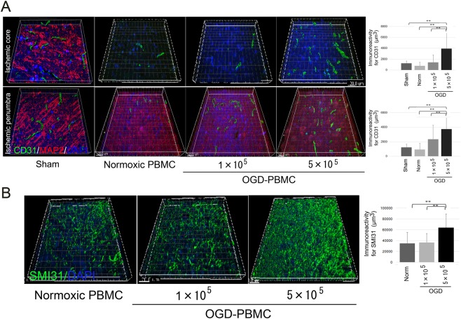
Figure 5.
OGD-PBMCs administration promotes both angiogenesis in the border area between the ischemic core and the penumbra and axonal outgrowth in the ischemic penumbra at 28 days after cerebral ischemia. (A) Representative figures and bar graphs representing the immunoreactivity for cluster of differentiation 31 (CD31) volume, expressed as μm3, in the ischemic core and penumbra from the cerebral cortices of the sham-operated group, and administration of the normoxic PBMCs group, 1 × 10⁵ OGD-PBMC, or 5 × 10⁵ OGD-PBMC groups at 28 days after cerebral ischemia. The CD31 (green)/MAP2 (red)/DAPI (blue) triple labelling in the ischemic cortices at 28 days after cerebral ischemia, as examined by confocal microscopy. (B) Representative figures and bar graphs representing immunoreactivity for SMI31-positive volumes, expressed as μm3, in the ischemic penumbra from the cerebral cortices of the administration of the normoxic PBMCs, 1 × 10⁵ OGD-PBMCs, or 5 × 10⁵ OGD-PBMCs groups. SMI31 (green)/DAPI (blue) double labelling in the ischemic cortices at 28 days after cerebral ischemia, as examined by confocal microscopy (N = 28). The bar graph represents the signal volume intensities of CD31 and SMI31 of brain samples by one-way ANOVA. Moreover, a secondary-only antibody control confirmed its specificity. Scale bars, 20 μm. **P < 0.01.

