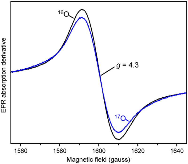
Figure 7.
EPR spectra of a 40 s sample of a single-turnover reaction of stoichiometrically reduced S5HH (472 μM) reacted with 16O2 (black, 720 μM) or 17O2 (blue, 676 μM, 50 % 17O2) and salicylate (8.88 mM) in standard buffer at 4 °C (concentrations after mixing). EPR spectra were normalized to the double integrated peak area of the g = 4.3 signal. Instrument conditions: microwave power, 200 μW; temperature, 2 K; modulation amplitude, 3 G; microwave frequency, 9.64 GHz.

